100 Years of ImagingHOME
|
|
Roentgen
(Written in 1995)
The forerunners
This year marks the centenary of the discovery of X rays. Some discoveries are the product of a sudden insight and some require extensive experimentation.
Newton's alleged encounter with the apple and his subsequent explanation of gravity is of the former type but Einstein required particles moving at the speed of light to produce the conditions necessary for him to arrive at the theory of relativity.
The discovery of X rays was somewhere in between. It required fairly sophisticated equipment but this equipment was commonly available. Some effects of X rays had already been observed but their significance had not been appreciated and it took Roentgen's inquiring mind to realise he was dealing with something new.
The equipment required to produce X rays was the cathode ray tube that was not very different in 1895 from the tube in your television set today.
The eccentric British scientist Crooke perfected the cathode ray tube. He frequently sent back photographic plates to the manufacturers complaining that light appeared to have got at them despite their light proof packages. The real explanation was that X rays from his own equipment were passing through the packaging.
In 1890 an American scientist Prof. Goodspeed of Philadelphia placed some coins on top of photographic plates in a drawer in his lab. After taking a photograph later with this plate he was surprised to see a double exposure of the coins as well as his own subject. Did he investigate - no he didn't - he put the photograph back in the drawer.
Later he sent it to Roentgen as the first X ray photograph but made no claim to the discovery of X rays. One person did make a claim to be the true discoverer and was declared as such many years later by Hitler (whom he just happened to support politically). His name was Prof. Lenard. Actually he had shown that cathode rays can be detected for a very short distance outside the tube and it was this experiment that Roentgen was repeating when he made his momentous discovery.
Mention must also be made of reports of a mysterious A. M. Esseltya, an Italian, who in 1848 was said to have demonstrated a machine that could take pictures of the inside of bodies. Sadly, no firm details are known.
Following Roentgen's discovery it became known that one of the assistants in his own lab had previously observed the same phenomenon as Roentgen but had failed to appreciate its significance. Roentgen
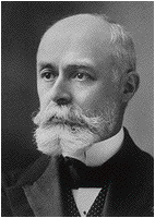
Conrad Roentgen was born in Lennep, Germany on 29th March 1845. As a young child his family moved to Appeldoorn in Holland to avoid political problems. Although a bright student, he ran into trouble at school having fallen foul of master for refusing to name fellow student who had performed some misdemeanour. He eventually gained a place at Zurich Polytechnic in Switzerland where he was nicknamed Apeldoorn.
He was an outstanding student but no bookworm. He became well known in Zurich due to his habit of dressing up and driving a fast carriage rather recklessly around the streets - perhaps the equivalent of a young leather clad student on a motorbike today. His other interests were opera, theatre, fencing and above all mountain climbing at which he excelled.
He enjoyed a litre or two of lager especially in the Green Tumbler in Zurich and later married Anna Bertha the daughter of the landlord, a man with a story to tell himself as he had only just escaped from helping to organise an attempted revolution in Jena in 1830. The marriage was successful but childless as Anna was tuberculous.
He married in 1870 having gained a post as an assistant in Wartburg. Later he worked in Strasburg then became a professor of physics in the University of Giessen where he gained a reputation as a first class research scientist.
In 1894 the University of Wartburg offered him the chair of physics and the directorship of physics department. He was nominated rector of the university in 1894/95 and only managed to work effectively towards the end of 1895.
On Friday 8th November he was trying to confirm Prof Lenard's finding that cathode rays could be identified for a very short distance outside the tube.
First he created a vacuum in a cathode ray tube surrounded with thin black card so that no light could escape. On switching on the tube he became aware of light coming from a table some distance away. The light proved to be coming from a screen of barium platinocyanide crystals. Barium platinocyanide is fluorescent which means it glows when light falls on it. But this glow could not be due to light because of the black card around the tube. Neither was it due to cathode rays as they only travel for a tiny distance.
Roentgen realised immediately that he had hit on something new. Being of Teutonic rather than Mediterranean origin he did not run down the streets of Wartburg shouting "Eureka!" but started immediately to investigate in a methodical way, although a friend did come across him in the street deep in thought and unable to recognise anybody. How did he investigate? His first experiment was to take the screen away some distance from the tube - the glow was weaker but still there. He placed a large book in front of the screen and it still glowed. These rays were passing through two hundred pages and the hardbacks. He found that metal stopped the rays. This led him to use a box of weights. There was the outline of the box but also the shadow of the weights. These rays were capable of producing a photograph of something inside a wooden box! Then he put his hand in front of the screen. He had already worked out what he would see but it was still quite a shock.
In a more sombre mood he then set about enumerating the properties of his new rays. As he did not know what they were he called them X Rays. A month later he produced a paper called "On a new kind of Ray" in which he outlined the basic physical properties of X rays: 1. They travel in a straight line 2. They penetrate materials to a degree depending on density and thickness. This paper contained an X Ray of his wife's hand.
Roentgen rapidly became famous, which he did not enjoy very much. He won the Nobel Prize in 1905. In 1922, still able to go on long walks at 77, he visited his beloved Swiss mountains and called in at the Green Tumbler. He died of an intestinal tumour on the10th February 1923 having worked almost to the end.
Early days of X Rays and the discovery of radioactivity
Immediately following Roentgen's discovery, the scientific world was understandably incredulous and disbelieving but the idea of invisible rays, which could pass through matter and take photos was an immediate hit with the general public. Fairground shows showing off the new rays sprang up everywhere. Some of these were genuine but mostly it was all done with mirrors. One firm in London produced X ray proof underwear. A law was passed in New York prohibiting the use of X ray opera glasses and one newspaper claimed that the new rays were being used to project anatomical details straight into the brains of medical students. Some very beautiful and informative pictures of both inanimate and animate objects were produced.
The ancestor of all diagnostic radiologists is a Dr Konig who in February 1896 undertook the first known medically useful X ray examination. A young boy had a swelling in his leg which was thought was probably a tumour. In those days the only treatment was amputation and surgeons were understandably reluctant to perform this operation. The X ray pictures were considered to be suggestive of malignancy and the amputation was carried out.
Radiology was taken up by doctors in various fields but many remained unconvinced and actively opposed its introduction, e.g. orthopaedic surgeons whose manipulation of fractures were shown to be not quite so straight as they claimed them to be and chest physicians, whose mastery of the stethoscope was being challenged. Similar attitudes were occasionally observed during the more recent introduction of ultrasound and MRI.
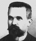
A stream of electrons alighting on a metal produces X-rays. Some scientists wondered if X-rays could be produced by incident light rather like the fluorescence mentioned above. Becquerel in Paris tried this theory out with various substances and soon found that uranium appeared to do just that. He exposed some uranium to light and then put it in a light proof compartment with a photographic plate and found that the plate was darkened. He tried to find a numerical association between the exposure to light and the X-ray energy given off but soon realised that the uranium was giving off its own rays. Radiation had been discovered. His discovery had little impact at first but then came the tragic figure of Marie Curie.
Born Marie Sklodowska of Polish parentage, she has a hard childhood. She married her tutor at the Sorbonne University, Pierre Curie, who, with his brother Jacques, was then living in near poverty in Paris. She discovered other radioactive substances and eventually realised that there was an element present in tiny amounts that was very powerfully radioactive and she set out to find it. She told Pierre that she wanted it to be pretty. They found a source of pitchblende waste from a mine and one ton of the unpleasant substance arrived from Austria on a cart.
The Curies started to extract chemically and physically the element that they knew was there and had already christened radium. Pierre worked at the University and Marie performed much of the hard physical work in extremes of cold and heat in her back yard for a period of four years. Eventually she obtained one decigram of radium. Was it pretty? It was more than pretty. Her daughter described how she remembers her parents holding hands in the back shed just watching their 1 dcgm of radium gently glowing blue in the dark.
Very soon they were famous; they had grants galore for their research and they could have been rich had they so wished. That was 1901. In 1903 Marie and Pierre were given a Nobel Prize jointly with Becquerel. In 1906 Pierre was run over by a cart in Paris and died. Marie was 38 and had two young daughters. She carried on with her work and did much useful radiology during the lst World War. She died in 1934 of aplastic anaemia, her blood cells having given up after so much irradiation.
Clinical Radiology
The two divisions of radiology: diagnostic and therapeutic were originally practised as one speciality. Oncologists went on to utilise other methods of treatment as well as radiation and after the Second World War diagnostic imaging began to encompass other forms of imaging.
There was some disagreement in the very early days as to whether specialist doctors or physicists should be providing an X ray service but the greater clinical input from the doctors proved decisive.
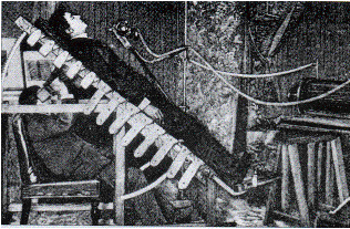
Early X ray machines were huge affairs involving Van der Graff generators, much noise and large sparks flying across the room. The X ray images were viewed on large fluorescent screens rather than photographs. A graphic description and an example of the philosophical deliberations that the images inspired can be found in Thomas Mann's book, "The Magic Mountain".
Rather surprisingly, it took quite a number of years before X-ray diagnosis became established. One doctor who did more than anyone to further the cause of diagnostic imaging was the Parisian physician Antoine Beclere who was already well on his way to a brilliant career in immunology having already developed a curative serum for smallpox. He became interested in radiology and realised that he could see far more on the X-ray screen if he allowed his eyes to become accustomed to the dark. Later he made an important contribution to ophthalmology by describing the biochemical changes that occur during dark adaptation. Because he spent so much time in the dark and began to see things that his less patient colleagues could not he began to get a reputation as an eccentric and perhaps a charlatan. He was a family man with a beard and his daughter describes how when he came to give her a goodnight kiss sparks would fly from his beard full of static.
He plodded on with his work and after many years gradually became more accepted by his colleagues. In 1906 came the case that turned the tide for diagnostic radiology. A young girl with pain in her upper right abdomen came into the hospital. She was very ill but in those days an abdominal operation was a hazardous undertaking and surgeons were reluctant to operate as the pain was in the wrong place for appendicitis. Beclere performed the equivalent of a barium meal and claimed that the patient did indeed have appendicitis but that, unusually, the appendix and the caecum were high up in the abdomen behind the liver.
One surgeon who had some faith in Beclere said he would operate if Beclere would sign a paper saying that he took full responsibility for what happened. Clearly the fear of medical litigation is not new. Using a much higher incision than usual, the operation was performed in an old fashioned theatre with a number of visiting surgeons watching in the gallery. Beclere was proved to be correct and the patient survived. These events were widely and internationally reported and Beclere's reputation soared. His ideas on light adaptation became widely practised, funds were allocated to X ray departments and the future of the speciality of radiology became assured. Beclere continued his work. He described the method of irradiating tumours from different angles to avoid skin burns and achieved a number of first time cures for cancers.
Other developments in Imaging include: 1. Angiography The injection of dense fluid into arteries in order to demonstrate them radiographically. The first angiograms were performed by Monitz in Italy in the 50s. His initial experiments were carried out a single dog. After he moved on to genuine patients he was surprised to find that the dog had become devoted to him and it became his pet, accompanying him to work every day.
2. Ultrasound was developed for diagnostic purposes also in the early 50s from wartime sonar. Special mention must be made of Prof. Donaldson at Glasgow, an obstetrician who produced the first useful pictures of fetuses.
3. Computerised Tomography (CT) and Magnetic Resonance Imaging (MRI) are both British inventions. Hounsfield, a physicist, produced the first CT Images at EMI and the teams at: Aberdeen, Sheffield and Nottingham developed MRI for medical use.
4. Interventional Radiology is the development of radiological techniques for treatment, e.g. the use of arteriographic techniques to unblock diseased arteries.
The Downside
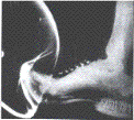
When Becquerel put some uranium in his pocket he noted that his skin was itching. Very early on it was noted that some radiologists began to get burns on their hands. These burns stayed for a long time and often forever. Lister, the grand old man of surgery, warned in 1896 that X-rays might be dangerous but not until 1912 did the real danger become apparent. Radiologists and their assistants were beginning to look ill. This was often put down to their working conditions: long hours in dark, damp cellars etc. But in 1912 an autopsy done on a Dr Tiraboschii in Italy found him to be anaemic with severe multiple organ damage and it was not long before the full dangers and the carcinogenic potential of X-Rays were realised.
There is an X-ray martyr's monument in Hamburg bearing 220 names of known victims of work induced radiation disease. As well as Radiologists the names include nurses and technicians.
Most martyrs to Roentgen's discovery are not on this monument. They are to be found in the records of Nagasaki and Hiroshima. One wonders what Becquerel would have done with his first specimen of uranium, already beginning to burn a warning mark on his chest from his jacket pocket as he walked to the Sorbonne. Would he have thrown it in the Seine had he been able to foresee the uses to which his discovery would be put? And what of gentle Roentgen; would it have been better for all of us if he had quietly turned his screen of platinocyanide round to face the wall?
|
History of radiology
Royal College of Radiologists
British Society of Interventional Radiologists
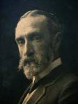
William Crooke
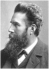
Conrad Roentgen
Henri Becquerel
Antoine Beclere
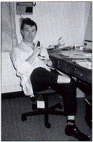
(and socks)
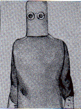
Early X-Ray protection
|